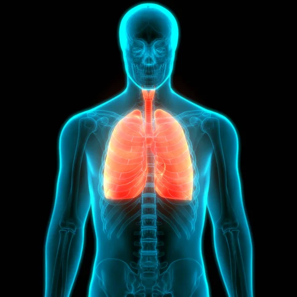
Anatomy Of Ribs And Lungs / Diaphragm Muscle Its Attachments And Actions Yoganatomy : Lungs are a pair of respiratory organs situated in a thoracic cavity.
Anatomy Of Ribs And Lungs / Diaphragm Muscle Its Attachments And Actions Yoganatomy : Lungs are a pair of respiratory organs situated in a thoracic cavity.. Anatomy of lungs lungs are a pair of respiratory organs situated in thoracic cavity. The infant has several developmental differences in the structure and function of the lung. The anatomy of bird's respiratory system, showing. They also have a role in ventilation; Lungs are known to be one of the most important organs as it facilitates respiration.
This begins at a somewhat higher level posteriorly, between the third and fifth ribs and runs downward and forward to end in the region of the sixth or seventh costochondral junction. Related online courses on physioplus. Anatomy of lungs lungs are a pair of respiratory organs situated in thoracic cavity. Neurons in this brain region send signals to the diaphragm and the muscles between the ribs to regulate the contractions which initiate the breathing. This page is about anatomy of ribs lungs and diaphragm,contains normal anatomy and flow during the complete examination:
Human Respiratory System Lungs Anatomy Stock Images Page Everypixel from st3.depositphotos.com The vessels canyon around the borders of the lung and margins of the fissures to reach the hilum. Includes images, video, and free quiz. Lungs are a pair of respiratory organs situated in a thoracic cavity. Occupying most of the space within the thoracic cavity, the lungs extend laterally from the heart to the ribs on both sides of the chest the bronchioles further branch off into many tiny terminal bronchioles. The arrangement of the air sacs, and lungs in birds. However the ribs decline inferiorly as they move around the thorax. The lungs are the primary organs of the respiratory system in humans and many other animals including a few fish and some snails. Neurons in this brain region send signals to the diaphragm and the muscles between the ribs to regulate the contractions which initiate the breathing.
Vestibular anatomy and neurophysiology online course:
Adults take 15 to 20 breaths a minute, which lungs do not have muscles to pump air in and out, though. The infant has several developmental differences in the structure and function of the lung. Vestibular anatomy and neurophysiology online course: The anterior border of the lung is formed by the convergence of the mediastinal. They are two in number, placed one on either side within the thorax, and separated from each other by the heart and other contents of the mediastinum (fig. The mediastinum, the cavity containing the heart, separates the two lungs. The lungs lie either side of the mediastinum, within the thoracic cavity. Vestibular anatomy and neurophysiology review the human postural control system to understand the impact of concussion. The mediastinal surface lies against the mediastinum anteriorly. As part of the bony thorax, the ribs protect the internal thoracic organs. They also have a role in ventilation; The main function of the lungs is to transport oxygen through the bloodstream to different parts of the body. Extracardiac anatomy,basics of pediatric anesthesia,the diaphragm,endometriosis & the heart and more.
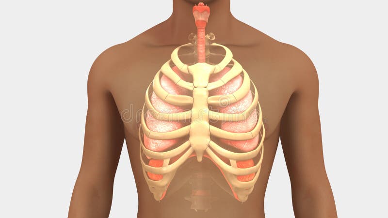
The heart and lungs work together to provide oxygen to the cells of the body. The rib cage is formed by the vertebral column, ribs, and sternum and encompasses the heart and lungs. Let's take a look at some anatomy of the lungs. Vestibular anatomy and neurophysiology review the human postural control system to understand the impact of concussion. This begins at a somewhat higher level posteriorly, between the third and fifth ribs and runs downward and forward to end in the region of the sixth or seventh costochondral junction.
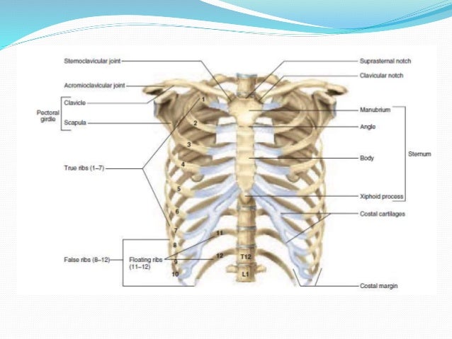
Lungs And Rib Cage Stock Illustration Illustration Of Throat 101914158 from thumbs.dreamstime.com The infant has several developmental differences in the structure and function of the lung. Terminal bronchioles are the smallest air tubes in the lungs and terminate at the. Includes images, video, and free quiz. Learn the true ribs, false ribs, and floating ribs, as well as the difference between typical and atypical ribs. The mediastinal surface lies against the mediastinum anteriorly. The anatomy of bird's respiratory system, showing. Anatomy of lungs lungs are a pair of respiratory organs situated in thoracic cavity. Vestibular anatomy and neurophysiology online course:
Lung anatomy pulmonary arteries and veins lung lobes.
Lungs are the vital organ of the respiratory system. The diaphragm and rib cage essentially there are around 480 million alveoli in the human lungs, according to the department of anatomy of. The vessels canyon around the borders of the lung and margins of the fissures to reach the hilum. The heart and lungs work together to provide oxygen to the cells of the body. Blunt lies above the level of anterior end of 1st rib. The lungs are the essential organs of respiration; As part of the bony thorax, the ribs protect the internal thoracic organs. Neurons in this brain region send signals to the diaphragm and the muscles between the ribs to regulate the contractions which initiate the breathing. Adults take 15 to 20 breaths a minute, which lungs do not have muscles to pump air in and out, though. They are also the ones responsible for releasing carbon. They also have a role in ventilation; ▪ introduction the lungs incorporate the parenchyma, vasculature, bronchial tree (trachea, bronchi, and bronchioles), and a network of investing connective tissue that supports and connects the structures the costal surface is the outer smooth and convex surface, which faces the ribs and the vertebrae. The rib cage is formed by the vertebral column, ribs, and sternum and encompasses the heart and lungs.
They are two in number, placed one on either side within the thorax, and separated from each other by the heart and other contents of the mediastinum (fig. Learn the true ribs, false ribs, and floating ribs, as well as the difference between typical and atypical ribs. This begins at a somewhat higher level posteriorly, between the third and fifth ribs and runs downward and forward to end in the region of the sixth or seventh costochondral junction. The pleura is a double‐layered membrane consisting of an inner pulmonary (visceral) pleura, which surrounds each lung, and an outer parietal pleura. However the ribs decline inferiorly as they move around the thorax.
Radiological Anatomy Of Chest Including Lungs Mediastinum And Thoraci from image.slidesharecdn.com Vestibular anatomy and neurophysiology review the human postural control system to understand the impact of concussion. Occupying most of the space within the thoracic cavity, the lungs extend laterally from the heart to the ribs on both sides of the chest the bronchioles further branch off into many tiny terminal bronchioles. Explore more like lungs and ribs anatomy. Extracardiac anatomy,basics of pediatric anesthesia,the diaphragm,endometriosis & the heart and more. However the ribs decline inferiorly as they move around the thorax. The ribs help protect vital organs in the thorax such as the heart and lungs, and they assist with breathing. Function of lungs and lung anatomy and lung lobes. While these anatomical variations are common and often go unnoticed in otherwise healthy individuals, it's important to distinguish them when reading.
The anterior, lateral, and posterior lung surfaces lie adjacent to the ribs and are thus often referred to as the costal surface.
Lungs are known to be one of the most important organs as it facilitates respiration. What is lung nodule, common lung disease & lung infection. Lung anatomy pulmonary arteries and veins lung lobes. Blunt lies above the level of anterior end of 1st rib. There are actually two lungs located in the chest. Lungs anatomy being demonstrated by showwing anatomical landmarks and surfaces of the lungs, in this interactive tutorial through labeled illustration. The anterior border of the lung is formed by the convergence of the mediastinal. Terminal bronchioles are the smallest air tubes in the lungs and terminate at the. The pleura is a double‐layered membrane consisting of an inner pulmonary (visceral) pleura, which surrounds each lung, and an outer parietal pleura. The lungs are a pair of cone‐shaped bodies that occupy the thorax. They are also the ones responsible for releasing carbon. They also have a role in ventilation; The costal surface of the lung this surface is large, smooth, and convex.
The arrangement of the air sacs, and lungs in birds anatomy of ribs. Function of lungs and lung anatomy and lung lobes.


0 Komentar